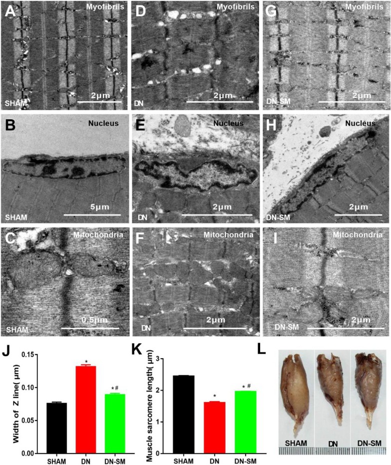
Fig. 2. Microstructure of gastrocnemius muscle fibers. (A-I) Transmission electron micrographs of longitudinal sections of the rat gastrocnemius muscle. Ultrastructure of skeletal myofibrils (A, D, G), nucleus (B, E, H), and mitochondria (C, F, I), width of the Z line (J), and the muscle sarcomere length (K) in the three groups (n = 3). (L) Representative images of the gastrocnemius muscle in the three groups.